Helpful Articles

Helpful Articles

Helpful Articles

Helpful Articles


It is common knowledge that exercise plays a critical role in healthy living. Exercise can help improve energy levels, lower blood pressure, aid in weight loss, helps to build muscle and so much more. But one benefit of exercise that is not as well known is the profound impact on your eyesight.
Based on recent research, eye conditions are usually a direct result of a health issue such as high blood pressure, diabetes, high cholesterol level, etc. While some of these diseases are unavoidable, exercising regularly can definitely help in the prevention of these diseases and in doing so, help keep the eyes healthy. In addition to physical exercise, there are even some eye exercises that can be done to keep your vision healthy. Some examples are focusing on certain points, rolling your eyes in different directions, writing with the eyes, etc.
Cataracts and Exercise
According to a study in 2003 and another in 2006, a relationship was discovered between an increase in exercise and a decrease in cataract. It emphasized that there is a greater chance for cataract if there is an absence of physical activity. This implies that taking a light walk or jog around your house or on a field track consistently can contribute to the fight against cataracts.
Macular Degeneration and Exercise
According to one study, exercising three times or more on a weekly basis reduces ones’ chances of developing exudative macular degeneration. Exudative macular degeneration occurs when blood vessels grow beneath the retina that are not needed and then leak fluid and blood into the eyes.
Glaucoma and Exercise
One major cause in the development of glaucoma is an increase in ocular perfusion pressure. Very light exercises like jogging or walking at least three times weekly helps to reduce the intraocular pressure and improves the flow of blood to the optic nerve and retina.
The benefit of eye exercises
Protects against dry eyes. Eye exercises help maintain and improve the fluid level in the eyes, thus eliminating the probability of dry eyes because as the eyes are being exercised say by rotation, there is the tendency that fluid is produced.
Reduces eye strain and tension. Exercising the eyes daily helps in reducing the strain and tension that has been placed on the muscle. Exercises like rolling the eyes and writing with the eyes help in the contracting and relaxing of the eye muscles. Long hours of study or use of a computer can cause blurry vision, but through exercise, clarity can be achieved by eliminating tension and unnecessary pressure.
Make the eye muscles flexible. Exercises like shifting and swinging the muscles of the eyes help promote flexibility in the muscles of the eyes. Just like exercise helps the body muscles, the same thing applies to eye exercises.
You might be surprised at how many tests eye doctors use to diagnose glaucoma. A proper diagnosis requires careful evaluation of many aspects of your eye’s health – from eye pressure to cornea thickness to the health of your optic nerve. This article describes how your eye doctor assesses your risk and all the tests needed to properly diagnose glaucoma.
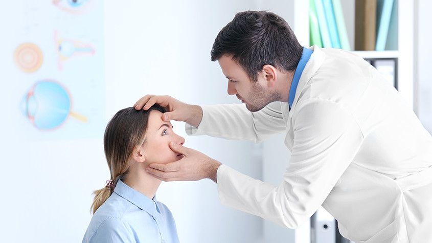
Risk Factor Assessment
Your eye doctor will begin by assessing your risk level for developing glaucoma. This will help determine the frequency and extent of testing needed. Through a family history and medical questionnaire, the eye doctor is looking for the following risk factors:
Over the age of 60
Ethnic background such as African or black Caribbean descent, Hispanic, or Asian
Family history of glaucoma, such as a sibling or parent with glaucoma
History of eye conditions, injuries or surgeries
Prolonged corticosteroid use (eye drops, pills, inhalers or creams)
Chronic conditions that affect blood flow, such as migraines, diabetes, low blood
pressure or hypertensionCurrent or former smoker
If you’ve already had a comprehensive eye exam, your eye doctor will also consider these risk factors:
Eye pressure higher than normal (above 21 mm Hg)
Thin corneas (less than 0.5 millimeters)
Your type of eyesight is also important. People with farsightedness are at a higher risk for narrow-angle glaucoma, a more serious type that can advance quickly. While nearsightedness is associated with open-angle glaucoma, which progresses slowly without any symptoms.
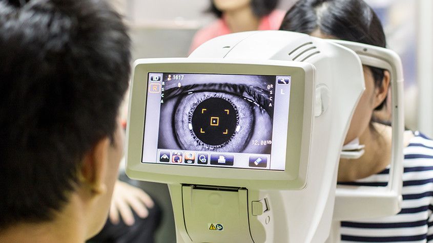
Standard Glaucoma Tests
During a comprehensive eye exam, your eye doctor will always check for glaucoma, regardless of the risk level. This provides a baseline for comparison as you age. There are two tests: tonometry and ophthalmoscopy.
Millions of patients are diagnosed with diseases and conditions of the eye every year. Some of which may not display symptoms until there is irreversible damage to the patient’s vision. The outcome of eye disease can range from temporary discomfort to total loss of vision, which is why all eye problems and diseases should be taken seriously and regular eye check-ups are absolutely essential.
What Are The Causes of Eye Disease?
The main causes of eye problems can be divided into five groups:
Inflammation of the eye and surrounding structures caused by a bacterial, viral, parasitic or fungal infection.
Injuries to the eye and surrounding structures, either as a result of trauma or an object in the eye.
Genetically inherited eye diseases, many of which may only manifest later in life and affect the structures and the functioning of the eye which therefore can impair visual abilities. In some cases, however, children are born with these conditions.
Diseases or conditions, such as migraine or diabetes, which can affect other organs of the body, such as the eyes.
External causes, such as allergies or eye strain due to over-use, or as a side effect of medication.
What Are The Symptoms of Eye Disease?
The three symptoms indicative of eye disease are changes in vision, changes in the
appearance of the eye, or an abnormal sensation or pain in the eye.
Changes in vision can include the following symptoms :
Nearsightedness is caused by an elongation of the eyeball over time, making it difficult to clearly see objects far away.
Farsightedness is caused by the shortening of the eyeball, making it difficult to see objects that are close-by clearly.
Blurry or hazy vision, or loss of specific areas of vision, which can affect one or both eyes and is the most common vision symptom. Any sudden changes in vision should be a cause of concern.
Double vision means a single clear image appears to repeat itself. This could be accompanied by other symptoms like headaches, nausea, a droopy eyelid, and misalignment of the eyes.
Floaters are specks or strands that seem to float across the field of vision. These are shadows cast by cells inside the clear fluid that fills the eye. These are usually harmless,

Generally, despite knowing how important the role of our eyes is in our day to day functioning, we tend to neglect the day to day care necessary to maintain optimal eye health. Just close your eyes for a minute or you put a non-transparent cloth across your eyes and try to walk around your home or workspace. It is quite difficult, right? That is how indispensable the eyes are.
While it is common knowledge that eye issues are a result of eyestrain or aging, most people are ignorant of the fact that an unbalanced diet, as well as deficiency in certain nutrients, can also facilitate eye issues. This implies that eating healthy means that there are certain nutrients that can help reduce the risk of eye issues. Some of these nutrients include omega-3 fatty acids, lutein, copper, zeaxanthin, zinc, vitamin E, A, C, and beta-carotene. For healthy eyes, there are foods that should be part of your diet daily. They include:
1. Fish- Fish is very rich in omega-3 fatty acids, especially oily fish which have oil inside their body tissue and guts. They are regarded as the major source of omega-3 fatty acids which help in improving the immune system and brain function. More so, it helps in the development of the eye and retina and in keeping the eyes from dryness. It is therefore important to incorporate fish into your diet about 3 times a week. Incorporating fish rich with this needed nutrient such as salmon, tuna, sardines, trout, herring, mackerel, and anchovies about 3 times a week is important in helping to maintain optimal eye health.
2. Eggs- The yolk of an egg contains a combination of the nutrients vitamin A, zeaxanthin, and lutein which help to safeguard the cornea, reducing the chances of suffering from cataracts and macular degeneration and zinc which keeps the retina healthy and aids night vision. Eggs are a very complimentary food as it can go with other foods and can also take various forms based on your taste.
3. Carrots- This is the most common food for eye health. It contains beta-carotene and vitamin A which protect the surface of the eyes and help to prevent infections. Vitamin A is a form of protein known as rhodopsin and it is tasked with the responsibility of aiding the eyes in absorbing light. A lack of vitamin A is the reason why there are about 500 thousand blind children every year. Carrots are easy to get and very affordable. They can serve as snacks or be sliced or diced into salads.
4. Nuts and Legumes- Legumes and nuts provide a lot of vitamin E and omega-3 fatty acids which guard the eyes against age-related disorders. Brazilian nuts, peanuts, walnuts, lentils, and cashews are perfect examples of legumes and nuts and can be eaten as a form of dessert or taken as snacks based on your choice.
5. Citrus Fruits- Citrus fruits are rich in vitamin C, an essential nutrient for healthy eyes. Vitamin C helps in keeping the blood vessels in the eyes healthy. It also stands against cataracts, and with other nutrients, it helps against macular degeneration. Citrus fruits include oranges, grapefruits, and lemons. The good thing about this food is that they can be consumed on their own or turned into a juice.

Eye disease that is caused by diabetes is currently the number one cause of blindness and vision loss. Due to the increased risk in diabetic patients, doctors recommend that people over 30 with diabetes get an annual dilated eye exam. Diabetic patients under 30 should get this exam five years after they have been diagnosed.
Diabetic retinopathy is a condition that is caused by damage to the retina. Patients that have diabetes may also have experienced extended periods of time where their blood sugar was elevated. The high levels of blood sugar damage the retina’s walls which leave them susceptible to leaking. When fluid accumulates in the retina or macula, it causes vision loss.
To make these matters worse, if prolonged high blood sugar levels are seen again, the retina will be oxygen-depleted. This causes the abnormal growth of new blood vessels. This condition is called neovascularization. This blood vessel type is weak and prone to leaking. As these blood vessels leak, they introduce blood into the eye. Excessive bleeding into the eye can cause blindness.
Treatment
While a healthy diet and exercise can be beneficial to your optical health, diabetic retinopathy is a condition that is caused by damage to the retinal wall. While this damage can sometimes be corrected, simple diet changes won’t reverse the effects.
It is essential to catch the condition in the earlier stages to reduce the effects. This can also help patients understand the importance of monitoring their blood sugar so that repeat events can be limited. Treatment options are even more successful when diabetic retinopathy is caught early. These options include vitrectomy, scatter photocoagulation and focal photocoagulation.
During both scatter, and focal photocoagulation ophthalmologists will use lasers to help alleviate the condition. The lasers make small burns on the retina aimed at the blood vessels. These burns will help to seal the blood vessels to prevent more leakage and stop them from growing larger.
When using scatter photocoagulation, hundreds of small burns are made in a specific pattern during two additional appointments. Scatter coagulation should be used on patients who do not have advanced diabetic retinopathy.
Focal photocoagulation specifically targets the leaking blood vessels that are in the macula. Unfortunately, this procedure is not aimed to correct the blurry vision associated with diabetic retinopathy, but it does stop it from progressing further. Once the retina has detached, neither form of photocoagulation can be used.
Vitrectomy is a surgery that helps to remove scar tissue and/or the fluid that is clouded with blood that has been leaked into the eye. This operation is the most successful when performed before the disease has progressed too far. When the operation only targets removing the fluid, success rates are very high for the procedure. When the procedure also aims to reattach the retina, the failure rate is around 50%.
Premium IOLs or intraocular lenses are lenses that are placed in the eye during cataract surgery. The lens placement is designed to restore the natural lens shape. These lenses can also be placed as a vision correction device called refractive lens exchange. Premium IOLs offer advanced features beyond the single vision IOL’s that are also offered. These features include aspheric, toric, accommodating, and multifocal IOL’s.
Understanding Premium IOL Types
Aspheric Lenses
These lenses closely match the natural curve of the eye. Typical lenses were uniformly curved making it easier to manufacture, but at the same time increasing the chance of causing imperfections in vision. Aspheric lenses help to reduce imperfections and improve clarity, especially at nighttime.
Toric IOLs
These lenses are specifically designed to help correct nearsightedness, farsightedness, and astigmatism.
Accommodating IOLs
Accommodating IOL’s can tilt slightly forward when you look at objects that are close to the eye. This helps to improve visibility when you are performing actions like reading a book. While they are not necessarily as sharp as bifocals, patients have a reduced need to use reading glasses while still maintaining excellent distance vision.
Multifocal IOLs
If you require a bifocal or trifocal lens in your glasses, this may be a likely choice for you. Different portions of the lens allow for better vision at different ranges. However, there are some overall sacrifices with vision clarity at a distance.
History of Premium IOLs
Premium IOLs have been approved by the Food and Drug Administration (FDA) since the 1980s. Prior to FDA approval, when patients had cataract surgery, they were required to wear very thick eyeglasses or specialized contact lenses to correct their vision. New technologies in the optical world have allowed for a wide variety of available premium IOLs and figuring out which specific type that suits you best will depend on some different factors.
Patient Factors
Physicians are careful to discuss the realities of this procedure with their patients. After recovering from cataract surgery, many patients expect that their vision will be completely restored to their peak performance. However, doctors are careful to warn against this and explain the realities of the surgery and as well as likely expectations of what will result from the surgery. For this reason, surgeons are likely to have some initial concern about the desired outcome for the patient to make sure that their hopes are grounded.
Surgeons will also have an eye toward the patient's desire to not wear eyeglasses. If the patient does not mind wearing corrective lenses without the need for surgery, this may be the best option. These lenses may also not be an ideal fit for the elderly population. Eyes in the geriatric population are often rapidly deteriorating requiring a lens replacement more quickly than would be recommended.
Patients with certain medical histories may also be poor candidates for premium IOL surgery. Some of these conditions include:
Advanced macular degeneration
Anterior basement membrane dystrophy
Fuch’s dystrophy
Weak zonules
Glaucoma
Post-refractive surgery patients
This list is not comprehensive, so it’s important to consult with your physician and bring a detailed medical history for their review.
Finally, patients may also want to consider their careers when weighing the value of this surgery. Patients who are required to read on computer screens for extended periods of time (i.e., print editors, office jobs) may be ideal candidates.
In contrast, individuals that require long-distance acuity like truck drivers, pilots, or even photographers may find that some of the issues with these lenses are not suited to their needs. Individuals often complain of “halos” during the night when looking toward a light, glare, or general acuity issues at longer distances.
Further Consideration
While premium IOLs do have some limitations, they offer an excellent choice for many individuals. However, it is important to meet with your eye care professional to fully discuss all of the available options to find your best fit as well as to make sure that you understand all of the potential risks and restrictions that this operation poses.
The parts of a comprehensive eye examination vary according to the patient's age, date of last exam and other factors. Not all parts of the eye exam may be needed or performed, but the first part of the eye exam will include documenting medical history. Here are some eye and vision tests that are likely to be encountered during a comprehensive eye exam:
Visual Acuity Tests
Visual acuity tests measure the sharpness of vision and are usually performed using a projected eye chart to measure the distance visual acuity and a hand-held small acuity chart to measure the near vision (for reading).
Color Blindness Test
A screening test that checks the color vision is often performed early in a comprehensive eye exam to rule out color blindness.
Cover test to check eye alignment.
A test used to assess strabismus or a more subtle binocular vision problem that could cause eye strain or amblyopia (lazy eye).
Ocular Motility (Eye Movements) Testing
Ocular motility testing is performed to determine how well eyes can follow a moving object and/or quickly move between and accurately fixate on two separate targets.
Stereopsis (Depth Perception) Test
This is used to test perception of depth and 3-dimensional structure obtained on the basis of visual information deriving from two eyes by individuals with normally developed binocular vision.
Retinoscopy
This test is used to estimate which lens powers will best correct distance vision. Based on the way the light reflects from the eye, the doctor is able to obtain an approximation of the eyeglass prescription. This test is useful for children and patients who are unable to accurately answer the doctor's questions.
Manual refraction with a phoropter.
This is the test used to determine the exact eyeglass prescription.

Understanding PRK: Is It Right for You?
PRK or photoreactive kerectomy is a surgical procedure that was the precursor for the surgery known as Lasik. The biggest difference between the two procedures is how the first portion of the operation is conducted. Additional variability between the two procedures includes recovery, risk factors, and the patient’s overall needs. Understanding these differences can help you decide if PRK is an appropriate solution for your vision issues.
How it Works
PRK utilizes a laser to correct farsightedness (hyperopia), nearsightedness (myopia), and astigmatism. During a PRK operation, a laser is used to remove the exterior epithelial cells from the cornea. This procedure uses an excimer laser to remove the cells which are then discarded. A contact “bandage” is placed over the eye, and the cells can heal over the course of a few days. Your doctor will then remove the contact lens when the eye has healed enough to be exposed.
While the results are like that of Lasik, PRK does take some additional healing time. This is due to the time that must be allowed for the epithelial cells to heal and regrow on the eye. Additionally, Lasik patients generally experience less discomfort and faster results. PRK results can take a few weeks to fully materialize.
This isn’t to say that PRK doesn’t have its own benefits too. This procedure is well-suited for patients that have had previous eye surgeries and may have thin corneas. Because PRK does not make an incision into the cornea and only removes the epithelial cells, it leaves more of the stromal tissues which underly the epithelial tissue. PRK does not run the risk of “flap” issues that can arise from Lasik, and the risk of removing too much of the cornea is reduced. However, if you are considering PRK, you should consult with your medical professional to identify the right procedure for your specific case.
Before the Surgery
When you meet with your eye specialist to discuss your options, there are several factors that they will consider. Your potential surgeon should conduct a thorough eye exam during which they will measure your eye moistness, pupil size, corneal thickness, and corneal curvature. Your doctor should also review your medical and family history to identify any possible concerns about your suitability. Make sure that you bring a list of your medications and previous operations. Finally, you may be required to stop wearing contact lenses for a period before the operation. This can allow your cornea to return to its natural shape before the operation.
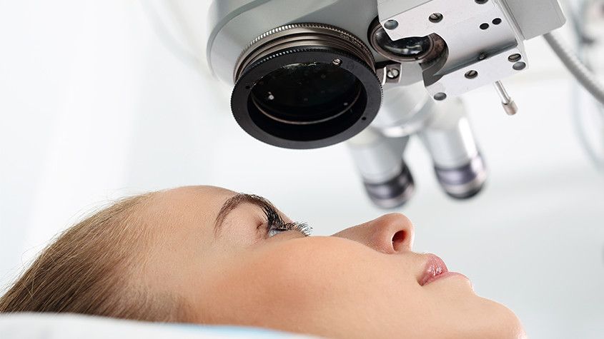
The Surgery
The actual PRK surgery is a short procedure that will only take about 15 minutes. The patient will not be sedated during the operation but may be given an oral sedative that helps to relax the eye. Numbing drops are applied to your eyes and a small speculum is also used to hold the eyelids open for the procedure. The excimer laser is programmed for your exact eye prescription. Patients are instructed to look at a certain object or target while the laser is operating. The surgeon will watch the procedure through a microscope and can stop the procedure at any time. Most patients do not report discomfort, although there may be some pressure.
Recovery
You will be observed for a short time after your operation to make sure that you don’t have any severe immediate reactions. After this observation, you will be sent home. It is important to have someone else drive you after any procedure that may impact your vision or ability to drive safely. You should make sure to follow all of the doctor’s recommendations to facilitate a speedy recovery. You should also expect several follow up appointments to make sure that the operation was successful and that there are no additional concerns.
Your full results may take several weeks, but almost all patients have vision that is 20/40 or better. Over time, as the eye ages, vision may naturally degrade. At this time, you should consult with your medical professional to see if an additional operation is a good option for you.
A refraction test, also called a vision test, is usually performed as a part of a routine eye examination. The purpose of this test is to determine if a person has a refractive error which would then mean the patient would need glasses or contact lenses.
What Is The Normal Value for Refraction Test?
A value of 20/20 is normal (optimum) vision. This means that individuals who have 20/20 vision are able to read letters that are 3/8-inch (1 centimeter) tall from 20 feet (6 meters) away. The normal uncorrected vision (without glasses or contact lenses) refractive error is zero (plano). Individuals who don’t have 20/20 vision, have what is called a refractive error. A refractive error means that the light is not bending properly when it passes through the lens of the eye. The refraction test will tell the doctor what prescription lens should be used in order to have 20/20 vision.
For people over age 40 who have normal distance vision but difficulty with near vision, a refraction test with a small type size is used to determine normal near vision and the correct power of reading glasses.
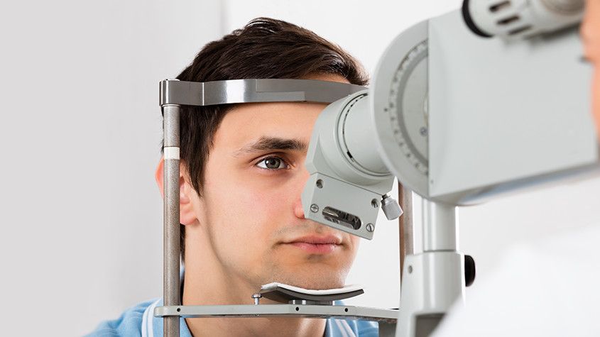
How Is The Refraction Test Performed?
The test is performed by having the patient seated in a chair that has a special device (called a phoropter or refractor) attached to it. The patient looks through the device and focuses on an eye chart 20 feet (6 meters) away. The device contains lenses of different strengths that can be moved into the patient’s view. The test is performed one eye at a time. If the patient is wearing contact lenses, they should be removed before the test.
In case the final vision is less than 20/20 even with lenses, then there is probably another non-optical problem with the eye. The vision level achieved during the refraction test is called the best-corrected visual acuity (BCVA).
What Are The Causes of Abnormal Refraction Test Results?
Abnormal results may be due to:
Astigmatism (abnormally curved cornea causing blurred vision)
Hyperopia (farsightedness)
Myopia (nearsightedness)
Presbyopia (inability to focus on near objects that develop with age)
Other conditions under which the test may be performed:
Corneal ulcers and infections
Loss of sharp vision due to macular degeneration
Retinal detachment (separation of the light-sensitive membrane (retina) in the back of the eye from its supporting layers)
Retinal vessel occlusion (blockage in a small artery that carries blood to the retina)
Retinitis pigmentosa (an inherited disorder of the retina)
There is an art to refraction and the ophthalmologists will always answer the patient’s questions and as well as discuss their findings. Based on the results of the refraction test, they can determine the amount of myopia, hyperopia or astigmatism.
If you’ve been diagnosed with glaucoma, you’re probably already familiar with the typical options in glaucoma treatment – eye drops, laser treatment or traditional surgery. While these are certainly effective, especially when glaucoma is diagnosed early, researchers have been working hard to offer new glaucoma treatments. Their goal is not only to improve outcomes but also reduce the treatment’s side effects and frequency of use.
What is the Goal of Glaucoma Treatment?
Before we dive into the new options, it’s important to understand the goal of any glaucoma treatment. At present, glaucoma is not curable. However, treatment can significantly slow the progression of the disease. Glaucoma damages your eye's optic nerve. Extra fluid builds up in the front part of your eye (cornea), which increases the pressure in your eye. Reducing this pressure is the primary objective of any glaucoma treatment. This is often referred to as intraocular pressure or IOP.
What are the Limitations of Standard Glaucoma Treatments?
Eye drops for glaucoma treatment seem like an easy option but there are several challenges that can reduce its effectiveness. It can be difficult to get all the medicine in the eye, especially for older adults with less of a steady hand. In addition, since it must be applied daily, individuals may forget. Since the drops have no perceivable benefit because early stages of glaucoma have no symptoms, patients might make it a lower priority which is understandable since it may also have unpleasant side effects like burning, red eyes.
Beyond eye drops, laser surgery is a less invasive option. The laser opens clogged tubes and drains fluid. It can take a few weeks to see the full results. If laser surgery or drugs don’t relieve your eye pressure, you may need a more traditional operation. You would have to go into the hospital and will need a few weeks to heal and recover. Although usually effective, glaucoma surgery can make you more likely to get cataracts later on. It can also cause eye pain or redness, infection, inflammation, or bleeding in your eye.
What are Recent Advances in Glaucoma Treatment?
Alternatives or Improvements to Eye Drops
The Glaucoma Research Foundation reported several new developments on the horizon. These technologies focus on reducing patient error in applying eye drops which would make the medication
Our Services
Contact Info
Hours of Operation
- Monday 9:00 AM - 6:00 PM
- Tuesday 9:00 AM - 6:00 PM
- Wednesday 9:00 AM - 6:00 PM
- Thursday 9:00 AM - 6:00 PM
- Friday 9:00 AM - 6:00 PM
- Saturday 9:00 AM - 1:00 PM
- Sunday Closed
- Closed for Lunch 1:00PM - 2:00PM
© 2024 Plano Eye Associates - All rights Reserved - Accessibility Statement - Sitemap
Powered by:
